- About
- Mission Statement
Education. Evidence. Regrowth.
- Education.
Prioritize knowledge. Make better choices.
- Evidence.
Sort good studies from the bad.
- Regrowth.
Get bigger hair gains.
Team MembersPhD's, resarchers, & consumer advocates.
- Rob English
Founder, researcher, & consumer advocate
- Research Team
Our team of PhD’s, researchers, & more
Editorial PolicyDiscover how we conduct our research.
ContactHave questions? Contact us.
Before-Afters- Transformation Photos
Our library of before-after photos.
- — Jenna, 31, U.S.A.
I have attached my before and afters of my progress since joining this group...
- — Tom, 30, U.K.
I’m convinced I’ve recovered to probably the hairline I had 3 years ago. Super stoked…
- — Rabih, 30’s, U.S.A.
My friends actually told me, “Your hairline improved. Your hair looks thicker...
- — RDB, 35, New York, U.S.A.
I also feel my hair has a different texture to it now…
- — Aayush, 20’s, Boston, MA
Firstly thank you for your work in this field. I am immensely grateful that...
- — Ben M., U.S.A
I just wanted to thank you for all your research, for introducing me to this method...
- — Raul, 50, Spain
To be honest I am having fun with all this and I still don’t know how much...
- — Lisa, 52, U.S.
I see a massive amount of regrowth that is all less than about 8 cm long...
Client Testimonials150+ member experiences.
 Scroll DownPopular Treatments
- Treatments
Popular treatments. But do they work?
- Finasteride
- Oral
- Topical
- Dutasteride
- Oral
- Topical
- Mesotherapy
- Minoxidil
- Oral
- Topical
- Ketoconazole
- Shampoo
- Topical
- Low-Level Laser Therapy
- Therapy
- Microneedling
- Therapy
- Platelet-Rich Plasma Therapy (PRP)
- Therapy
- Scalp Massages
- Therapy
 More
More
IngredientsTop-selling ingredients, quantified.
- Saw Palmetto
- Redensyl
- Melatonin
- Caffeine
- Biotin
- Rosemary Oil
- Lilac Stem Cells
- Hydrolyzed Wheat Protein
- Sodium Lauryl Sulfate
 More
More
ProductsThe truth about hair loss "best sellers".
- Minoxidil Tablets
Xyon Health
- Finasteride
Strut Health
- Hair Growth Supplements
Happy Head
- REVITA Tablets for Hair Growth Support
DS Laboratories
- FoliGROWTH Ultimate Hair Neutraceutical
Advanced Trichology
- Enhance Hair Density Serum
Fully Vital
- Topical Finasteride and Minoxidil
Xyon Health
- HairOmega Foaming Hair Growth Serum
DrFormulas
- Bio-Cleansing Shampoo
Revivogen MD
- more
Key MetricsStandardized rubrics to evaluate all treatments.
- Evidence Quality
Is this treatment well studied?
- Regrowth Potential
How much regrowth can you expect?
- Long-Term Viability
Is this treatment safe & sustainable?
Free Research- Free Resources
Apps, tools, guides, freebies, & more.
- Free CalculatorTopical Finasteride Calculator
- Free Interactive GuideInteractive Guide: What Causes Hair Loss?
- Free ResourceFree Guide: Standardized Scalp Massages
- Free Course7-Day Hair Loss Email Course
- Free DatabaseIngredients Database
- Free Interactive GuideInteractive Guide: Hair Loss Disorders
- Free DatabaseTreatment Guides
- Free Lab TestsProduct Lab Tests: Purity & Potency
- Free Video & Write-upEvidence Quality Masterclass
- Free Interactive GuideDermatology Appointment Guide
- More
Articles100+ free articles.
-
Can Miniaturized Hair Follicles Recover, or Is There a Point of No Return?
-
Sublingual Minoxidil: More Hair Growth, Less Sides?
-
Happy Head Review: 5 Concerns With The Brand
-
Can a Blood DHT Test Tell You Whether You Have AGA?
-
ABS-201: Can Blocking the Prolactin Receptor Regrow Hair?
-
Best Topical Finasteride: 5 Top Products of 2026
-
Best Topical Dutasteride: 8 Choices for 2026
-
2.5mg Dutasteride: Before and After Photos [2026]
PublicationsOur team’s peer-reviewed studies.
- Microneedling and Its Use in Hair Loss Disorders: A Systematic Review
- Use of Botulinum Toxin for Androgenic Alopecia: A Systematic Review
- Conflicting Reports Regarding the Histopathological Features of Androgenic Alopecia
- Self-Assessments of Standardized Scalp Massages for Androgenic Alopecia: Survey Results
- A Hypothetical Pathogenesis Model For Androgenic Alopecia:Clarifying The Dihydrotestosterone Paradox And Rate-Limiting Recovery Factors
Menu- AboutAbout
- Mission Statement
Education. Evidence. Regrowth.
- Team Members
PhD's, resarchers, & consumer advocates.
- Editorial Policy
Discover how we conduct our research.
- Contact
Have questions? Contact us.
- Before-Afters
Before-Afters- Transformation Photos
Our library of before-after photos.
- Client Testimonials
Read the experiences of members
Before-Afters/ Client Testimonials- Popular Treatments
-
ArticlesCan Miniaturized Hair Follicles Recover, or Is There a Point of No Return?
First Published Oct 13 2026Last Updated Oct 13 2026Natural RemediesPharmaceutical Researched & Written By:Sarah King, PhD
 Reviewed By:Perfect Hair Health Team
Want help with your hair regrowth journey?
Get personalized support, product recommendations, video calls, and more from our researchers, trichologists, and PhD's dedicated to getting you the best possible outcomes.
Learn MoreArticle Summary
In androgenic alopecia, hair follicles do not disappear overnight. Instead, they get progressively smaller, producing shorter, finer, less pigmented hairs that cannot cover the scalp. This article explains what happens to follicles in AGA, whether miniaturized hair follicles are “dead” or dormant, what treatments can realistically reverse, and why earlier treatment usually produces better outcomes.
Full Article
When someone notices a thinning crown, a widening part, or a receding hairline, it is natural to assume that the follicles in those areas are dying. But in androgenic alopecia (AGA), that is not usually what happens, at least not in the way most people mean it.
AGA is primarily a condition of hair follicle miniaturization. The follicle often remains in the scalp, but it gradually shrinks and produces lower-quality hair fibers. A thick, long, pigmented terminal hair might become a shorter, finer, less pigmented vellus hair that contributes very little to visible coverage.
This, therefore, raises the question: if a follicle has not simply disappeared, could it recover and return to its former state as a terminal hair?
The short answer is sometimes.
Treatments like finasteride and minoxidil can slow miniaturization and improve visible hair growth in many people. However, they must be used continuously to maintain their effects, and they do not reliably restore every follicle to the size or productivity it had before AGA began. Furthermore, while studies suggest that bald AGA scalps can retain some stem cell populations, this does not mean that all long-bald follicles are merely “asleep” and ready to be reactivated.
To understand why, we first need to look at how AGA affects the normal hair cycle.
What Happens to Hair Follicles in AGA?
A hair follicle is not a permanent, continuously growing structure. It repeatedly moves through a cycle of growth, regression, rest, and renewal.
The three main phases are:[1]Kloepper, J.E., Sugawara, K., Al-Nuaimi, Y., Gaspar, E., & Paus, R. (2010). Methods in hair research: How to objectively distinguish between anagen and catagen in human hair follicles. … Continue reading,[2]Alonso, L., & Fuchs, E. (2006). The hair cycle. Journal of Cell Science. 119(Pt 3). 391-393. Available at: https://doi.org/10.1242/jcs.02793
- Anagen: The active growth phase. The follicle produces a hair shaft, and scalp hairs can remain in anagen for years.
- Catagen: A brief regression phase. Growth stops, the lower part of the follicle shrinks, and the hair-producing bulb moves upward.
- Telogen: The resting phase. The follicle remains relatively inactive before eventually returning to anagen. The old club hair may shed during this period or shortly before the next hair begins to grow.
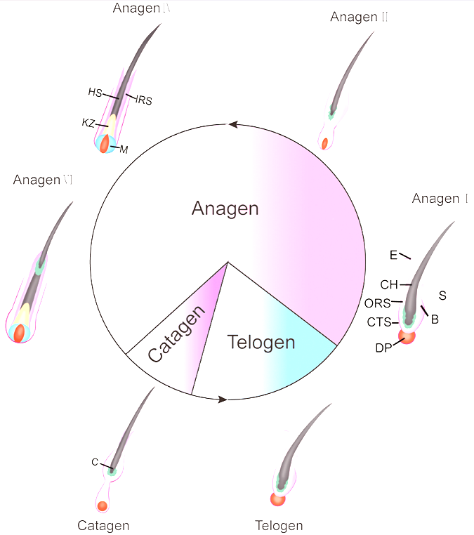
Figure 1: The human hair follicle cycle goes through growth (anagen), regression (catagen), and resting (telogen) phases.[3]Lin, X., Zhu, L., He, J. (2022). Morphogenesis, Growth Cycle and Molecular Regulation of Hair Follicles. Frontiers in Cell and Developmental Biology. 10(899095). Available at: … Continue reading Image used in conjunction with the PMC Copyright Notice.
In healthy scalp hair, anagen is unusually long. That is why terminal scalp hairs can grow to substantial lengths. But in AGA-prone areas of the scalp, the cycle gradually changes.
Genetically susceptible hair follicles respond to androgen signaling, particularly dihydrotestosterone (DHT), by producing hairs that are progressively smaller with each cyle. The anagen phase shortens, meaning each hair has less time to grow. At the same time, the follicle that is produced is thinner and finer.[4]Randall, V.A. (2008). Androgens and hair growth. Dermatologic Therapy. 21(5). 314-328. Available at: https://doi.org/10.1111/j.1529-8019.2008.00214.x,[5]Sawaya, M.E., & Price, V.H. (1997). Different levels of 5-alpha-reductase type I and II, aromatase, and androgen receptor in hair follicles of women and men with androgenetic alopecia. Journal of … Continue reading
This is called the terminal-to-vellus transition.
Terminal hairs are the thick, pigmented hairs that create most of the scalp’s visible coverage, whereas vellus hairs are much finer, shorter, and less pigmented. In AGA, a follicle may not abruptly become a true vellus follicle. Instead, it gradually moves along a spectrum, producing hairs that become increasingly vellus-like over repeated cycles.
In practical terms, AGA does not usually mean that a hair follicle simply “falls out”. It means that the follicle becomes smaller and less capable of making a cosmetically useful hair.
Why AGA Does Not Look Uniform
One reason AGA can be difficult to assess in its early stages is that follicles do not miniaturize at the same speed.
Two adjacent follicles can be at very different stages. One may still produce a thicker terminal hair, while the follicle beside it may produce a short, fine, miniaturized fiber. This produces a mixture of hair diameters within the same scalp area, a feature known as hair-diameter diversity, or anisotrichosis.
That mixture is one of the most useful signs of AGA under trichoscopy. Rather than a uniform reduction across all hairs, clinicians often observe increasing variation. Some hairs remain relatively thick, while others are much thinner and shorter. A 2024 cross-sectional study in men with AGA found that hair-diameter diversity increased with disease severity, while mean hair diameter declined and the proportion of thinner hairs rose.[6]Kim, M.S., Kim, J., Lee, S.Y., et al. (2024). Quantitative measurement of hair diameter diversity as a diagnostic indicator of androgenetic alopecia in Korean males: A cross-sectional study. JAAD … Continue reading
This is why AGA often looks like reduced density rather than immediate baldness. The hairs may still be present, but an increasing proportion are too fine to provide meaningful coverage.
Are Miniaturized Follicles Dead or Dormant?
The short answer is that neither word is completely accurate.
Calling a miniaturized AGA follicle “dead” suggests that it has been physically destroyed, which is simply not the case.
AGA is classified as a non-scarring alopecia. Even when an area of scalp looks bald, miniaturized follicular structures may still remain. This differs from scarring alopecia, where inflammation can physically destroy follicles and replace them with fibrotic tissue. In those conditions, follicular openings can disappear, and the potential for spontaneous regrowth is much more limited.[7]Vidal, C.I. (2015). Overview of alopecia: A dermatopathologist’s perspective. Missouri Medicine. 112(4). 308-312. Available at: https://pmc.ncbi.nlm.nih.gov/articles/PMC6170065/
But “dormant” is not quite right either.
A miniaturized AGA follicle may still cycle and produce a hair shaft. The issue is that the produced shaft may be too fine, short, or lightly pigmented to provide noticeable scalp coverage. In other words, it’s not necessarily inactive, but rather functioning at a reduced level.
It should be noted that AGA can still become difficult to reverse, especially in longstanding bald areas. But its biology is not identical to a scarring disorder. A miniaturized follicle and a physically destroyed follicle are not the same thing.
What Stem Cell Studies Actually Show
One influential study compared bald and haired scalps from men with AGA. Researchers found that bald scalp retained KRT15-positive cells associated with the hair follicle’s bulge region, a stem-cell niche located in the permanent portion of the hair follicle. However, they also found marked depletion of several additional activated progenitor-cell populations located near the bulge, including CD200-high/ITGA6-high and CD34-positive cells.[8]Garza, L.A., Yang, C.C., Zhao, T., et al. (2011). Bald scalp in men with androgenetic alopecia retains hair follicle stem cells but lacks CD200-rich and CD34-positive hair follicle progenitor cells. … Continue reading
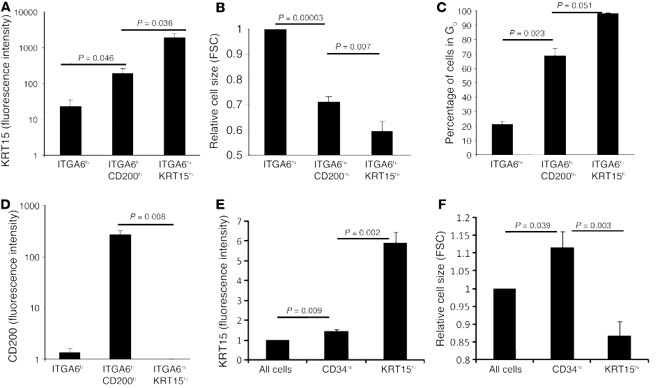
Figure 2: (A–C) CD200-high/ITGA6-high cells had intermediate levels of KRT15, cell size, and time spent in a resting, non-dividing state (G0) when compared with all basal-layer keratinocytes and the more quiescent KRT15-high stem-cell population. (D) Most KRT15-high cells had little or no CD200 expression, supporting the idea that they represent a distinct cell population. (E–F) CD34-high cells had lower KRT15 levels and were larger than KRT15-high cells. Together, these findings suggest that CD200-high/ITGA6-high and CD34-high cells differ from the KRT15-high stem-cell population and may represent more activated, progenitor-like stages of the hair-follicle cell lineage. FSC, forward scatter, a flow-cytometry measure used as an estimate of cell size.[9]Garza, L.A., Yang, C.C., Zhao, T., et al. (2011). Bald scalp in men with androgenetic alopecia retains hair follicle stem cells but lacks CD200-rich and CD34-positive hair follicle progenitor cells. … Continue reading
At first glance, that might sound like very good news. If stem cells are still there, perhaps the follicles can simply be switched back on.
But that conclusion goes beyond what the study showed.
A more cautious interpretation is that some of the hair follicle’s core machinery persists in the bald AGA scalp, while the processes required to activate those cells or convert them into the progenitor cells needed for robust hair production may be impaired.
Put differently, the issue may not be a total absence of follicular stem cells; it might be a failure of the signaling, activation, or stem-cell-to-progenitor conversion steps required to rebuild a normal terminal hair.
That is an important finding for regenerative hair loss research. It is not proof that every long-bald follicle can currently be restored with conventionally available treatments.
Can AGA Treatments Reverse Miniaturization?
The most accurate answer is: partially, in some follicles, in some people.
That may sound less exciting than claims of “follicle revival” or “complete regrowth”, but it reflects what the clinical evidence actually supports.
A positive response to treatment can include:
- A longer anagen phase, allowing hairs to grow for longer before entering regression.
- Increased hair-shaft diameter, making existing hairs more visible.
- A shift from producing very fine, vellus-like hairs toward producing thicker, more cosmetically meaningful hairs.
- Recruitment of follicles that were producing very little visible hair into a more productive growth cycle.
- Slower ongoing miniaturization, which can preserve density that would otherwise have been lost.
These changes can substantially improve scalp coverage. But they are not necessarily equivalent to full anatomical restoration of every affected follicle.
What Finasteride Studies Show
Finasteride reduces the conversion of testosterone to DHT by inhibiting type II 5-alpha-reductase. In men with AGA, this reduces DHT in serum and scalp tissue, thereby lowering one of the key androgenic signals that drives miniaturization in genetically susceptible follicles.[10]Drake, L., Hordinsky, M., Fiedler, V., et al. (1999). The effects of finasteride on scalp skin and serum androgen levels in men with androgenetic alopecia. Journal of the American Academy of … Continue reading
Randomized trials have shown that finasteride can increase hair counts and improve photographic appearance compared with placebo in men with AGA.[11]Kaufman, K.D., Olsen, E.A., Whiting, D., et al. (1998). Finasteride in the treatment of men with androgenetic alopecia. Journal of the American Academy of Dermatology. 39(4 Pt 1). 578-589. Available … Continue reading
Long-term evidence is particularly useful here because AGA is progressive. In a five-year multinational study, men taking finasteride 1 mg daily showed durable improvements in hair count, investigator assessments, patient assessments, and clinical photographs, while the placebo group continued to lose hair over time.[12]Shapiro, J., Kaufman, K.D., Olsen, E.A., Whiting, D., & Savin, R. (2003). Long-term (5-year) multinational experience with finasteride 1 mg in men with androgenetic alopecia. European Journal of … Continue reading
However, “finasteride works” should not be interpreted as “finasteride can restore your original hair density”.
Some men do see visible regrowth, while others maintain what they already have. Some improve in one area, like the crown, but not the frontal hairline. Some have a modest response despite a substantial reduction in DHT, while others do not experience meaningful cosmetic improvement.
This is because DHT reduction is only one part of the chain between drug exposure and visible hair growth. The final outcome depends on disease duration, degree of miniaturization, follicular biology, treatment adherence, age, genetics, and the amount of potentially recoverable hair remaining.
For many people, preventing further decline is not a treatment failure. In a progressive condition such as AGA, stabilizing hair loss can be a meaningful outcome.
What Does Minoxidil Do?
Minoxidil works through a different treatment pathway than finasteride. It does not meaningfully reduce DHT or remove the follicle’s underlying androgen sensitivity.
Instead, minoxidil appears to influence hair cycling and follicular productivity. Its exact mechanism is not fully understood, but research suggests that it can promote anagen activity, influence potassium channels and intracellular signaling, and affect growth-factor pathways within the follicular environment.[13]Messenger, A.G., & Rundegren, J. (2004). Minoxidil: Mechanisms of action on hair growth. British Journal of Dermatology. 150(2). 186-194. Available at: … Continue reading,[14]Suchonwanit, P., Thammarucha, S., & Leerunyakul, K. (2019). Minoxidil and its use in hair disorders: A review. Drug Design, Development and Therapy. 13. 2777-2786. Available at: … Continue reading
Clinically, minoxidil can increase total and non-vellus hair growth compared with a placebo. A systematic review and meta-analysis reported improvements in total and non-vellus hair counts with topical minoxidil.[15]Fiedler, V.C., Alaitz, M., & Hayden, R.E. (2015). Topical minoxidil: Systematic review and meta-analysis of its efficacy in androgenetic alopecia. Journal of the American Academy of Dermatology. … Continue reading
In a 48-week trial in men with AGA, 5% topical minoxidil outperformed both 2% minoxidil and placebo for non-vellus hair count, investigator-rated scalp coverage, and patient-rated treatment benefit.[16]Olsen, E.A., Dunlap, F.E., Funicella, T., et al. (2002). A randomized clinical trial of 5% topical minoxidil versus 2% topical minoxidil and placebo in the treatment of androgenetic alopecia in men. … Continue reading
The important limitation is that minoxidil does not eliminate the underlying androgen sensitivity of AGA. That is why its benefits are generally treatment-dependent. If treatment is stopped, the hair that was being maintained or improved by minoxidil may gradually be lost.
Why Combination Therapy Can Help
Studies and reviews of combination minoxidil and finasteride treatment generally suggest better outcomes than monotherapy, at least for some patients.[17]Gupta, A.K., Bamimore, M.A., & Foley, K.A. (2020). The efficacy and safety of finasteride combined with topical minoxidil for androgenetic alopecia: A systematic review and meta-analysis. Journal … Continue reading Using both makes biological sense. One treatment can slow the process driving miniaturization, while the other may improve the productivity of the remaining follicles.
What About Microneedling, PRP, and Experimental Treatments?
Microneedling, platelet-rich plasma (PRP), low-level light therapy (LLLT), topical anti-androgens, and cell-based or regenerative approaches are frequently discussed in the treatment of AGA.
Some of these interventions have encouraging signals.
For example, systematic reviews of PRP have found that PRP injections can improve hair density compared with control treatments in several small randomized trials. However, PRP preparation, platelet concentration, injection technique, treatment schedule, comparator treatment, and outcome measurement vary substantially between studies.[18]Zhang, X., Ji, Y., Zhou, M., et al. (2023). Platelet-rich plasma for androgenetic alopecia: A systematic review and meta-analysis of randomized controlled trials. Journal of Cutaneous Medicine and … Continue reading
Microneedling combined with minoxidil has also shown better average hair-count outcomes than minoxidil alone in meta-analyses. But the evidence remains heterogeneous, with studies using different needle depths, devices, treatment intervals, and follow-up periods.[19]Zhou, Y., Wang, J., Liu, Y., et al. (2025). Evaluating the efficacy and safety of combined microneedling with minoxidil versus minoxidil monotherapy for androgenetic alopecia: A systematic review and … Continue reading
The key issue is not whether these interventions can improve average hair-count measurements. Some certainly do. The question is whether those changes demonstrate anatomical reversal of miniaturization in longstanding follicles.
Right now, the answer is no.
Prolactin-Receptor Inhibitors
While the above treatments can be effective, they aren’t designed specifically to address the possibility that AGA involves defective communication between hair-follicle stem cells and the progenitor cells needed to build a robust terminal hair.
That is one reason prolactin-receptor inhibitors have attracted attention.
The prolactin receptor is expressed in hair follicles, and experimental work suggests that prolactin signaling can influence hair-cycle regulation. New injectable antibodies, like Absci’s ABS-201, are being developed to block prolactin receptor signaling in people with AGA.
The appeal of this approach is straightforward. If miniaturized follicles retain some stem-cell populations but fail to generate enough active progenitor cells, a treatment that improves the follicle’s regenerative environment could, in theory, help more follicles return to productive growth.
But “in theory” is the important phrase here.
HMI-115, which we have covered in more detail here, is one of the first PRLR-blocking treatments to reach early-stage clinical testing for AGA. In a company-reported open-label Phase 1b study, 12 male participants with AGA reportedly had a mean increase of 14 non-vellus hairs per cm2 after 24 weeks of treatment.[20]Hope Medicine. (2024). Positive outcome from a Phase Ib study in Australia treating androgenetic alopecia with HMI-115. Company press release. Available at: … Continue reading This result is promising, but the study was small, lacked a placebo comparison, and has not yet provided the kind of peer-reviewed, full clinical dataset needed to determine how reliably HMI-115 improves hair density, caliber, or long-term outcomes.
ABS-201 is an anti-prolactin receptor antibody in the earlier stages of clinical development. Its ongoing Phase 1/2a HEADLINE trial is randomized, double-blind, and placebo-controlled, with planned measures including target-area hair count, hair width, and pigmentation.[21]Absci. (2026). Absci announces positive interim Phase 1 data from the HEADLINE trial evaluating ABS-201 in androgenetic alopecia. Company press release. Available at: … Continue reading However, the company’s initial human update focused on safety and pharmacokinetics in the single-ascending-dose portion of the trial, rather than confirmed hair-growth efficacy.
Absci has also reported ex vivo findings from human scalp tissue suggesting that PRLR blockade may prolong anagen and improve growth-associated follicular activity.[22]Absci. (2025). Absci reports new human ex vivo data demonstrating that ABS-201 promotes hair follicle growth. Company press release. Available at: … Continue reading
For now, prolactin-receptor inhibitors are best viewed as an intriguing test of a newer biological hypothesis: that improving the regenerative signaling environment of the follicle may complement conventional DHT-lowering and growth-promoting therapies. Whether this translates into clinically meaningful reversal of advanced miniaturization remains unknown.
Why Earlier Treatment Usually Produces Better Results
Early intervention tends to be more rewarding because, even if follicles are starting to produce finer, shorter hairs, they may still have greater regenerative capacity than follicles further along in the process.
There are two potential benefits to treating earlier:
- Preservation: Slowing miniaturization before thicker terminal hairs become very fine or disappear from view.
- Thickening: Improving the caliber, length, and visibility of hairs produced by follicles that remain viable but are already underperforming.
This is why preventing further deterioration can be a successful outcome even without dramatic regrowth.
Someone who began treatment while they still had a dense field of miniaturized hairs may see a substantial cosmetic improvement if enough of those hairs thicken. Someone with a longstanding slick-bald area may have fewer follicles capable of producing visible hair, even if some follicular structures remain in the scalp.
Does Fibrosis Create a Point of No Return?
Perifollicular inflammation and fibrosis have been observed in AGA, particularly around the upper follicle. Histological studies have identified inflammatory cell infiltrates and fibrotic changes in at least some of the affected scalp areas.[23]Jaworsky, C., Kligman, A.M., Murphy, G.F., & Kennedy, J.W. (1992). Characterization of inflammatory infiltrates in male pattern alopecia: Implications for pathogenesis. British Journal of … Continue reading,[24]Mahé, Y.F., Michelet, J.F., Billoni, N., et al. (2000). Androgenetic alopecia and microinflammation. International Journal of Dermatology. 39(8). 576-584. Available at: … Continue reading
This has led to an understandable theory. Perhaps fibrosis gradually locks a follicle into a permanent non-responsive state.
But the evidence is not strong enough to say that fibrosis is the sole cause of irreversible AGA.
AGA is a complex condition involving several factors, one of which may be fibrosis, but it should not be treated as the sole explanation for why recovery becomes harder.
The Arrector Pili Muscle Hypothesis
Another proposed explanation involves the arrector pili muscle, the tiny smooth muscle attached to the hair follicle that causes goosebumps.
Research suggests that miniaturized hair follicles in AGA may lose their normal attachment to the arrector pili muscle. This is notable because miniaturized follicles in alopecia areata may preserve this attachment and can often regrow terminal hair when the autoimmune process resolves.[25]Yazdabadi, A., Whiting, D., Rufaut, N., & Sinclair, R. (2012). Miniaturized hairs maintain contact with the arrector pili muscle in alopecia areata but not in androgenetic alopecia. International … Continue reading
This has been proposed as one reason why AGA miniaturization may become harder to reverse than miniaturization in alopecia areata. However, this is best understood as a biological model rather than a clinically validated “point-of-no-return” test. No clinician can currently examine a scalp and use arrector pili attachment alone to calculate the chance that a particular follicle will regrow.
How Can You Estimate Whether a Hair Follicle Might Respond?
There is a non-invasive test that can prove whether a specific follicle is recoverable.
But clinicians can look for clues to help estimate whether an area of the scalp still contains follicles capable of producing more visible hair.
Visible Miniaturized Hairs
Fine, short hairs in an area of thinning are often more encouraging than completely smooth, long-bald skin. While this does not guarantee regrowth, it suggests that follicles in the area are still producing shafts, even if those shafts are currently too fine to provide useful density.
Hair-Shaft Diameter Diversity
A mixture of thick terminal hairs and fine miniaturized hairs is a hallmark of active AGA miniaturization. Trichoscopy can show this variation more clearly than the naked eye. It can also identify an increased proportion of vellus-like hairs, changes in follicular units, and regional density differences.[26]Kuczara, A., Waskiel-Burnat, A., Rakowska, A., Olszewska, M., & Rudnicka, L. (2024). Trichoscopy of androgenetic alopecia: A systematic review. Journal of Clinical Medicine. 13(7). 1962. … Continue reading
Density Under Standardized Trichoscopy
A standardized trichoscopic examination or phototrichogram can help document:
- Hair density per square centimeter.
- Average shaft diameter.
- The proportion of terminal versus vellus-like hair.
- Changes in the same scalp area over time.
Duration and Speed of Progression
Duration matters, but it is not a precise predictor.
An area that has been slick-bald for many years is generally less likely to respond than an area that has recently thinned and still contains visible miniaturized hairs. But there is no scientifically validated rule stating that follicles become irreversibly “dead” after a specific number of years.
Speed also matters. Rapid recent thinning may warrant evaluation for additional contributors, such as telogen effluvium, inflammatory scalp disease, medication effects, nutritional deficiency, or endocrine conditions.
Duration and Speed of Progression
Duration matters, but it is not a precise predictor.
An area that has been slick-bald for many years is generally less likely to respond than an area that has recently thinned and still contains visible miniaturized hairs. But there is no scientifically validated rule stating that follicles become irreversibly “dead” after a specific number of years.
Speed also matters. Rapid recent thinning may warrant evaluation for additional contributors, such as telogen effluvium, inflammatory scalp disease, medication effects, nutritional deficiency, or endocrine conditions.
Response to an Adequate Treatment Trial
Ultimately, an individual’s response after a sufficiently long and consistent treatment trial is often more useful than a baseline prediction.
AGA treatments work slowly because hair cycling is slow. A short trial of a few weeks or a couple of months is generally not enough to judge whether miniaturized hairs are thickening or whether progression has stabilized.
Photographs can help, but only if they are standardized.
For useful comparisons, take photographs with the same:
- Lighting
- Camera distance
- Camera angle
- Hair length
- Hairstyle or part placement
- Scalp dryness and hair product use
- Head position
Phone photographs alone cannot determine whether follicles are viable. They can show changes in coverage, but they cannot distinguish miniaturization from shedding, breakage, changes in hair length, or lighting changes.
A scalp biopsy is also not normally required solely to predict regrowth in typical AGA. Biopsy is generally reserved for uncertain cases, atypical presentations, or situations where scarring alopecia or another diagnosis needs to be excluded.
Is There a Proven Point of No Return?
No.
There is currently no validated hair-diameter threshold, duration of baldness, Norwood stage, fibrosis score, or trichoscopic measurement that defines the exact point at which an AGA follicle becomes irreversibly non-responsive.
Longstanding bald areas are less likely to produce substantial regrowth with existing medical treatments than areas containing numerous fine hairs. But less likely is not the same as impossible, and current evidence does not allow clinicians to calculate a precise individual probability from duration alone.
This uncertainty can be frustrating. But it is better than false certainty in either direction.
Final Thoughts
AGA miniaturization is best understood as a continuum rather than an on/off process. Affected follicles do not usually vanish all at once; instead, they gradually become smaller and produce hairs that are shorter, finer, less pigmented, and less able to cover the scalp. Because neighboring follicles can be at different stages of miniaturization, AGA often creates a mixture of thick terminal hairs and fine vellus-like hairs rather than uniform hair loss. This is also why “dead” and “dormant” are imperfect descriptions: a miniaturized follicle may still be present and may still produce a hair shaft, but it may no longer produce hair that contributes meaningfully to cosmetic density.
Research suggesting that some follicular stem-cell populations persist in bald AGA scalp is biologically important, but it should not be interpreted as proof that long-bald follicles can currently be restored to their original state. The more realistic takeaway is that treatment can sometimes reverse part of miniaturization, particularly in follicles that are still producing fine, visible hairs. Finasteride, minoxidil, and combination approaches may help preserve existing hair, slow further diameter loss, and improve the thickness, length, or visibility of hair that remains responsive. This is why earlier treatment generally offers the greatest opportunity: the goal is not always dramatic regrowth or a return to original density, but preventing further deterioration while improving the follicles that are still capable of producing visible hair.
References[+]
References ↑1 Kloepper, J.E., Sugawara, K., Al-Nuaimi, Y., Gaspar, E., & Paus, R. (2010). Methods in hair research: How to objectively distinguish between anagen and catagen in human hair follicles. Experimental Dermatology. 19(4). 305-312. Available at: https://doi.org/10.1111/j.1600-0625.2009.00939.x ↑2 Alonso, L., & Fuchs, E. (2006). The hair cycle. Journal of Cell Science. 119(Pt 3). 391-393. Available at: https://doi.org/10.1242/jcs.02793 ↑3 Lin, X., Zhu, L., He, J. (2022). Morphogenesis, Growth Cycle and Molecular Regulation of Hair Follicles. Frontiers in Cell and Developmental Biology. 10(899095). Available at: https://doi.org/10.3389/fcell.2022.899095 ↑4 Randall, V.A. (2008). Androgens and hair growth. Dermatologic Therapy. 21(5). 314-328. Available at: https://doi.org/10.1111/j.1529-8019.2008.00214.x ↑5 Sawaya, M.E., & Price, V.H. (1997). Different levels of 5-alpha-reductase type I and II, aromatase, and androgen receptor in hair follicles of women and men with androgenetic alopecia. Journal of Investigative Dermatology. 109(3). 296-300. Available at: https://doi.org/10.1111/1523-1747.ep12335779 ↑6 Kim, M.S., Kim, J., Lee, S.Y., et al. (2024). Quantitative measurement of hair diameter diversity as a diagnostic indicator of androgenetic alopecia in Korean males: A cross-sectional study. JAAD International. 15. 121-126. Available at: https://doi.org/10.1016/j.jdin.2024.02.005 ↑7 Vidal, C.I. (2015). Overview of alopecia: A dermatopathologist’s perspective. Missouri Medicine. 112(4). 308-312. Available at: https://pmc.ncbi.nlm.nih.gov/articles/PMC6170065/ ↑8, ↑9 Garza, L.A., Yang, C.C., Zhao, T., et al. (2011). Bald scalp in men with androgenetic alopecia retains hair follicle stem cells but lacks CD200-rich and CD34-positive hair follicle progenitor cells. Journal of Clinical Investigation. 121(2). 613-622. Available at: https://doi.org/10.1172/JCI44478 ↑10 Drake, L., Hordinsky, M., Fiedler, V., et al. (1999). The effects of finasteride on scalp skin and serum androgen levels in men with androgenetic alopecia. Journal of the American Academy of Dermatology. 41(4). 550-554. Available at: https://pubmed.ncbi.nlm.nih.gov/10495374/ ↑11 Kaufman, K.D., Olsen, E.A., Whiting, D., et al. (1998). Finasteride in the treatment of men with androgenetic alopecia. Journal of the American Academy of Dermatology. 39(4 Pt 1). 578-589. Available at: https://doi.org/10.1016/s0190-9622(98)70007-6 ↑12 Shapiro, J., Kaufman, K.D., Olsen, E.A., Whiting, D., & Savin, R. (2003). Long-term (5-year) multinational experience with finasteride 1 mg in men with androgenetic alopecia. European Journal of Dermatology. 13(6). 555-561. Available at: https://pubmed.ncbi.nlm.nih.gov/11809594/ ↑13 Messenger, A.G., & Rundegren, J. (2004). Minoxidil: Mechanisms of action on hair growth. British Journal of Dermatology. 150(2). 186-194. Available at: https://doi.org/10.1111/j.1365-2133.2004.05785.x ↑14 Suchonwanit, P., Thammarucha, S., & Leerunyakul, K. (2019). Minoxidil and its use in hair disorders: A review. Drug Design, Development and Therapy. 13. 2777-2786. Available at: https://doi.org/10.2147/DDDT.S214907 ↑15 Fiedler, V.C., Alaitz, M., & Hayden, R.E. (2015). Topical minoxidil: Systematic review and meta-analysis of its efficacy in androgenetic alopecia. Journal of the American Academy of Dermatology. 72(5). AB59. Available at: https://pubmed.ncbi.nlm.nih.gov/26380504/ ↑16 Olsen, E.A., Dunlap, F.E., Funicella, T., et al. (2002). A randomized clinical trial of 5% topical minoxidil versus 2% topical minoxidil and placebo in the treatment of androgenetic alopecia in men. Journal of the American Academy of Dermatology. 47(3). 377-385. Available at: https://pubmed.ncbi.nlm.nih.gov/12196747/ ↑17 Gupta, A.K., Bamimore, M.A., & Foley, K.A. (2020). The efficacy and safety of finasteride combined with topical minoxidil for androgenetic alopecia: A systematic review and meta-analysis. Journal of Cosmetic Dermatology. 19(12). 3219-3228. Available at: https://doi.org/10.1111/jocd.13658 ↑18 Zhang, X., Ji, Y., Zhou, M., et al. (2023). Platelet-rich plasma for androgenetic alopecia: A systematic review and meta-analysis of randomized controlled trials. Journal of Cutaneous Medicine and Surgery. 27(6). 620-630. Available at: https://doi.org/10.1177/12034754231191461 ↑19 Zhou, Y., Wang, J., Liu, Y., et al. (2025). Evaluating the efficacy and safety of combined microneedling with minoxidil versus minoxidil monotherapy for androgenetic alopecia: A systematic review and meta-analysis. Journal of Cosmetic Dermatology. Available at: https://pubmed.ncbi.nlm.nih.gov/40056230/ ↑20 Hope Medicine. (2024). Positive outcome from a Phase Ib study in Australia treating androgenetic alopecia with HMI-115. Company press release. Available at: https://www.hopemedinc.com/company-release-37 ↑21 Absci. (2026). Absci announces positive interim Phase 1 data from the HEADLINE trial evaluating ABS-201 in androgenetic alopecia. Company press release. Available at: https://investors.absci.com/news-releases/news-release-details/absci-announces-positive-interim-phase-1-data-headlinetm-trial/ ↑22 Absci. (2025). Absci reports new human ex vivo data demonstrating that ABS-201 promotes hair follicle growth. Company press release. Available at: https://investors.absci.com/news-releases/news-release-details/absci-reports-new-human-ex-vivo-data-demonstrating-abs-201tm/ ↑23 Jaworsky, C., Kligman, A.M., Murphy, G.F., & Kennedy, J.W. (1992). Characterization of inflammatory infiltrates in male pattern alopecia: Implications for pathogenesis. British Journal of Dermatology. 127(3). 239-246. Available at: https://doi.org/10.1111/j.1365-2133.1992.tb00121.x ↑24 Mahé, Y.F., Michelet, J.F., Billoni, N., et al. (2000). Androgenetic alopecia and microinflammation. International Journal of Dermatology. 39(8). 576-584. Available at: https://doi.org/10.1046/j.1365-4362.2000.00612.x ↑25 Yazdabadi, A., Whiting, D., Rufaut, N., & Sinclair, R. (2012). Miniaturized hairs maintain contact with the arrector pili muscle in alopecia areata but not in androgenetic alopecia. International Journal of Trichology. 4(1). 28-31. Available at: https://pmc.ncbi.nlm.nih.gov/articles/PMC3500053/ ↑26 Kuczara, A., Waskiel-Burnat, A., Rakowska, A., Olszewska, M., & Rudnicka, L. (2024). Trichoscopy of androgenetic alopecia: A systematic review. Journal of Clinical Medicine. 13(7). 1962. Available at: https://doi.org/10.3390/jcm13071962 Want help with your hair regrowth journey?
Get personalized support, product recommendations, video calls, and more from our researchers, trichologists, and PhD's dedicated to getting you the best possible outcomes.
Learn More
Sarah King, PhD
Dr. Sarah King is a researcher & writer who holds a BSc in Medical Biology, an MSc in Forensic Biology, and a Ph.D. in Molecular and Cellular Biology. While at university, Dr. King’s research focused on cellular aging and senescence through NAD-dependent signaling – along with research into prostaglandins and their role in hair loss. She is a co-author on several upcoming manuscripts with the Perfect Hair Health team.
"... Can’t thank @Rob (PHH) and @sanderson17 enough for allowing me to understand a bit what was going on with me and why all these [things were] happening ... "

 — RDB, 35, New York, U.S.A.
"... There is a lot improvement that I am seeing and my scalp feel alive nowadays... Thanks everyone. "

 — Aayush, 20’s, Boston, MA
"... I can say that my hair volume/thickness is about 30% more than it was when I first started."

 — Douglas, 50’s, Montréal, Canada
Want help with your hair regrowth journey?
Get personalized support, product recommendations, video calls, and more from our researchers, trichologists, and PhD's dedicated to getting you the best possible outcomes.
Join Now - Mission Statement