- About
- Mission Statement
Education. Evidence. Regrowth.
- Education.
Prioritize knowledge. Make better choices.
- Evidence.
Sort good studies from the bad.
- Regrowth.
Get bigger hair gains.
Team MembersPhD's, resarchers, & consumer advocates.
- Rob English
Founder, researcher, & consumer advocate
- Research Team
Our team of PhD’s, researchers, & more
Editorial PolicyDiscover how we conduct our research.
ContactHave questions? Contact us.
Before-Afters- Transformation Photos
Our library of before-after photos.
- — Jenna, 31, U.S.A.
I have attached my before and afters of my progress since joining this group...
- — Tom, 30, U.K.
I’m convinced I’ve recovered to probably the hairline I had 3 years ago. Super stoked…
- — Rabih, 30’s, U.S.A.
My friends actually told me, “Your hairline improved. Your hair looks thicker...
- — RDB, 35, New York, U.S.A.
I also feel my hair has a different texture to it now…
- — Aayush, 20’s, Boston, MA
Firstly thank you for your work in this field. I am immensely grateful that...
- — Ben M., U.S.A
I just wanted to thank you for all your research, for introducing me to this method...
- — Raul, 50, Spain
To be honest I am having fun with all this and I still don’t know how much...
- — Lisa, 52, U.S.
I see a massive amount of regrowth that is all less than about 8 cm long...
Client Testimonials150+ member experiences.
 Scroll DownPopular Treatments
- Treatments
Popular treatments. But do they work?
- Finasteride
- Oral
- Topical
- Dutasteride
- Oral
- Topical
- Mesotherapy
- Minoxidil
- Oral
- Topical
- Ketoconazole
- Shampoo
- Topical
- Low-Level Laser Therapy
- Therapy
- Microneedling
- Therapy
- Platelet-Rich Plasma Therapy (PRP)
- Therapy
- Scalp Massages
- Therapy
 More
More
IngredientsTop-selling ingredients, quantified.
- Saw Palmetto
- Redensyl
- Melatonin
- Caffeine
- Biotin
- Rosemary Oil
- Lilac Stem Cells
- Hydrolyzed Wheat Protein
- Sodium Lauryl Sulfate
 More
More
ProductsThe truth about hair loss "best sellers".
- Minoxidil Tablets
Xyon Health
- Finasteride
Strut Health
- Hair Growth Supplements
Happy Head
- REVITA Tablets for Hair Growth Support
DS Laboratories
- FoliGROWTH Ultimate Hair Neutraceutical
Advanced Trichology
- Enhance Hair Density Serum
Fully Vital
- Topical Finasteride and Minoxidil
Xyon Health
- HairOmega Foaming Hair Growth Serum
DrFormulas
- Bio-Cleansing Shampoo
Revivogen MD
- more
Key MetricsStandardized rubrics to evaluate all treatments.
- Evidence Quality
Is this treatment well studied?
- Regrowth Potential
How much regrowth can you expect?
- Long-Term Viability
Is this treatment safe & sustainable?
Free Research- Free Resources
Apps, tools, guides, freebies, & more.
- Free CalculatorTopical Finasteride Calculator
- Free Interactive GuideInteractive Guide: What Causes Hair Loss?
- Free ResourceFree Guide: Standardized Scalp Massages
- Free Course7-Day Hair Loss Email Course
- Free DatabaseIngredients Database
- Free Interactive GuideInteractive Guide: Hair Loss Disorders
- Free DatabaseTreatment Guides
- Free Lab TestsProduct Lab Tests: Purity & Potency
- Free Video & Write-upEvidence Quality Masterclass
- Free Interactive GuideDermatology Appointment Guide
- More
Articles100+ free articles.
-
Can a Blood DHT Test Tell You Whether You Have AGA?
-
ABS-201: Can Blocking the Prolactin Receptor Regrow Hair?
-
Best Topical Finasteride: 5 Top Products of 2026
-
Best Topical Dutasteride: 8 Choices for 2026
-
2.5mg Dutasteride: Before and After Photos [2026]
-
Best Minoxidil for Women: Top 7 Brands of 2026
-
Pumpkin Seed Oil for Hair Growth: Before and After Photos
-
Best Minoxidil for Men: Top Picks for 2026
PublicationsOur team’s peer-reviewed studies.
- Microneedling and Its Use in Hair Loss Disorders: A Systematic Review
- Use of Botulinum Toxin for Androgenic Alopecia: A Systematic Review
- Conflicting Reports Regarding the Histopathological Features of Androgenic Alopecia
- Self-Assessments of Standardized Scalp Massages for Androgenic Alopecia: Survey Results
- A Hypothetical Pathogenesis Model For Androgenic Alopecia:Clarifying The Dihydrotestosterone Paradox And Rate-Limiting Recovery Factors
Menu- AboutAbout
- Mission Statement
Education. Evidence. Regrowth.
- Team Members
PhD's, resarchers, & consumer advocates.
- Editorial Policy
Discover how we conduct our research.
- Contact
Have questions? Contact us.
- Before-Afters
Before-Afters- Transformation Photos
Our library of before-after photos.
- Client Testimonials
Read the experiences of members
Before-Afters/ Client Testimonials- Popular Treatments
-
ArticlesCan a Blood DHT Test Tell You Whether You Have AGA?
First Published Oct 7 2026Last Updated Oct 2 2026PharmaceuticalUncategorized Researched & Written By:Sarah King, PhD
 Reviewed By:Rob English, Medical Editor
Want help with your hair regrowth journey?
Get personalized support, product recommendations, video calls, and more from our researchers, trichologists, and PhD's dedicated to getting you the best possible outcomes.
Learn MoreArticle Summary
A blood DHT test may seem like a clear way to explain hair thinning, but it cannot diagnose androgenic alopecia on its own. This article explains why scalp follicles respond to local androgen activity, genetic susceptibility, and follicle-specific signalingnot simply the amount of DHT circulating in the blood, and outlines when hormone testing may still be clinically useful.
Full Article
If you are noticing a receding hairline, widening part, reduced density, or progressively finer hairs, a blood DHT test can sound like a logical next step.
Dihydrotestosterone (DHT) is often described as the hormone responsible for androgenic alopecia (AGA). That framing can make the question seem straightforward. If DHT contributes to follicular miniaturization, wouldn’t a high DHT result show whether DHT is causing your hair loss?
There are other understandable reasons to want the test, too. You may be trying to distinguish AGA from stress-related shedding or a nutritional problem. You may want an objective explanation for a visible change in your hair. Or if you are considering a treatment such as finasteride, you may wonder whether your baseline DHT level could predict whether the medication will work for you.
A blood test can feel more definitive than looking at the scalp. It provides a number, a laboratory reference range, and apparently clear categories such as “normal” or “high.” But hair follicles do not respond only to the amount of DHT circulating in the bloodstream. They respond to a combination of local DHT production, androgen-receptor signaling, genetic susceptibility, and site-specific biology within the scalp.
That does not make serum DHT meaningless. It is a real hormonal measurement, and hormone testing can be appropriate when someone has signs of androgen excess or another possible endocrine disorder. However, interpreting a DHT result as a direct test for AGA can be misleading.
To understand why, it helps to separate two related but different questions:
- What is the DHT concentration in the blood at the time of testing?
- Are susceptible scalp follicles undergoing androgen-related miniaturization?
What is DHT?
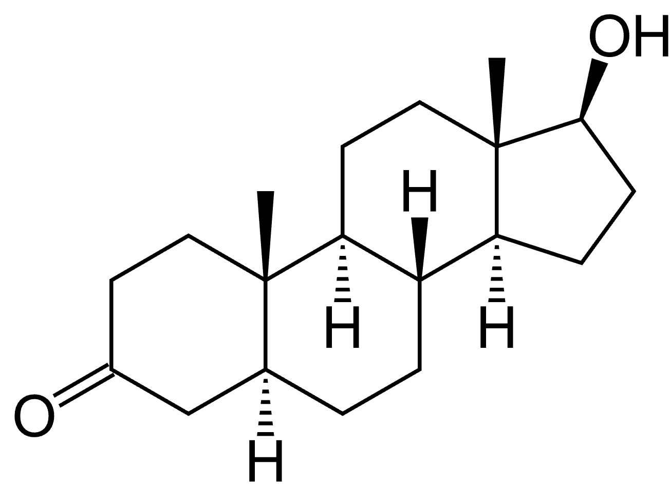
Dihydrotestosterone, or DHT, is an androgen hormone created from the conversion of testosterone by 5-alpha-reductase.[1]Kinter KJ, Amraei R, Anekar AA. Biochemistry, Dihydrotestosterone. [Updated 2023 Jul 30]. In: StatPearls [Internet]. Treasure Island (FL): StatPearls Publishing; 2026 Jan-. Available from: … Continue reading
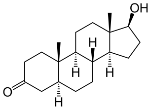
Dihydrotestosterone.[2]Wikimedia Commons. (no date). Dihydrotestosterone. Available at: https://commons.wikimedia.org/wiki/File:Androstanolone.svg (Accessed: September 2026)
DHT binds to the androgen receptor, a protein inside certain cells that acts as a molecular receiver for androgen signals. Once DHT binds, the androgen receptor can influence the expression of genes involved in tissue growth, development, and function.
There are several forms of 5-alpha-reductase. In hair biology, the two most often discussed are:
- Type I 5-alpha-reductase, which is found in skin and pilosebaceous structures, among other tissues.
- Type II 5-alpha-reductase, which is particularly relevant in androgen-sensitive tissues and is a key therapeutic target of finasteride.
Both enzymes can contribute to the local conversion of testosterone into DHT, but their distribution and relative importance vary by tissue, sex, and scalp location.
DHT is not inherently “bad”. It has important physiological roles, particularly in male sexual development and in androgen-dependent tissues. The apparent contradiction is that DHT can stimulate terminal hair growth in some areas, such as the beard, chest, and other body sites, while contributing to progressive miniaturization of susceptible scalp follicles.
This is not because beard and scalp follicles receive different blood DHT concentrations, but because follicles in different areas of the body interpret androgen signals differently. Their local enzyme activity, androgen-receptor expression, surrounding signaling environment, and genetic programming all differ.[3]Itami, S., Sonoda, T., Kurata, S., Takayasu, S. (1994). Mechanism of action of androgen in hair follicles. Journal of Dermatological Sciences. S98-103. Available at: … Continue reading,[4]Rutberg, S.E., Kolpack, M.L., Gourley, J.A., Tan, G., Henry, J.P., Shander, D. (2006). Differences in expression of specific biomarkers distinguish human beard from scalp dermal papilla cells. … Continue reading
How DHT contributes to AGA
AGA is best understood as an interaction among several factors:
- Androgen exposure, including testosterone and DHT
- Local production and metabolism of androgens within the scalp
- Activity of 5-alpha-reductase enzymes in and around the follicle
- Androgen receptor amount and responsiveness
- Genetic susceptibility
- Follicle-specific signaling pathways that influence hair-cycle duration, inflammation, growth factors, and miniaturization
In other words, AGA is not simply “high DHT”. It is a condition in which particular scalp follicles are susceptible to androgen-mediated change.
Human hair follicle research illustrates this point. In a study of frontal and occipital scalp follicles from men and women with AGA, Sawaya and Price found site- and sex-specific differences in levels of androgen receptors, type I and type II 5-alpha-reductase, and aromatase.[5]Sawaya, M.E., Price, V.H. (1997). Different levels of 5-alpha-reductase type I and II, aromatase, and androgen receptor in hair follicles of women and men with androgenetic alopecia. Journal of … Continue reading Frontal follicles had higher androgen-receptor and 5-alpha-reductase levels, while occipital follicles had higher aromatase levels. The differences were also quantitatively distinct between women and men.
This helps explain why hair loss often follows a pattern. In many men, the frontal scalp, temples, and vertex are more vulnerable than the occipital scalp. In many women, thinning is more apparent over the central scalp or widening part line, while the frontal hairline may be relatively preserved.
It also explains why balding and non-balding follicles can behave differently within the same person. Two follicles may be exposed to the same circulating DHT concentration, yet one remains stable while the other progressively miniaturizes.
Why the same DHT level can mean different things
Consider two people with the same serum DHT results:
- One may have follicles with relatively low local 5-alpha-reductase activity, lower androgen receptor signaling, or low genetic susceptibility to AGA.
- The other may have genetically susceptible frontal or vertex follicles with greater local androgen signaling and a stronger miniaturizing response
The blood test may look similar. The clinical hair outcome may be completely different.
That is why a serum result cannot substitute for assessment of the scalp itself.
What a blood DHT test measures
A blood DHT test measures the concentration of DHT in serum at one particular point in time. It does not directly measure what is occurring inside your scalp follicles.
A reported result can also be influenced by several factors:
- The laboratory method used to measure DHT
- The laboratory’s reference range
- Normal biological variation over time
- Timing of sampling
- Sex, age, and hormonal status
- Use of oral contraceptives, hormone therapy, anabolic-androgenic steroids, or anti-androgen medications
- Use of 5-alpha-reductase inhibitors such as finasteride or dutasteride
- Other medical conditions that influence androgen production, metabolism, or binding proteins
DHT measurement can be technically challenging, especially at the relatively low concentrations often found in women. Where hormone testing is clinically necessary, assay quality matters. For example, endocrine guidance commonly emphasizes high-quality testosterone measurement and assessment of related markers such as sex hormone-binding globulin (SHBG), rather than treating DHT as a standalone diagnostic marker.[6]Elhassan, Y.S., Hawley, J.M., Cussen, L., Abbara, A., Clarke, S.A., Kempegowda, P., Dhillon-Smith, R.K., Thadani, P., Busby, M., Owusu-Darkwah, L., Marrington, R., Duncan, C.W., Semple, R.K., … Continue reading
A blood DHT result does not tell you:
- The DHT concentration inside an individual scalp follicle
- Local 5-alpha-reductase activity in the balding scalp
- Androgen receptor density or sensitivity
- Genetic susceptibility to AGA
- Whether miniaturization is occurring
- How advanced miniaturization is
- Whether another type of hair loss is present instead
Serum and scalp DHT are related pharmacodynamic measures: medications that reduce DHT systemically can also reduce it in the scalp. But they are not interchangeable measures. A serum concentration is not a direct readout of follicular androgen signaling or a diagnostic marker of scalp miniaturization.
Do people with AGA have higher serum DHT?
The evidence is mixed. Some studies report group-level differences in circulating androgen markers between people with AGA and control participants. However, group differences do not automatically create a useful diagnostic test for an individual patient.
Research finding What it means clinically A group with AGA has a higher average DHT value than a control group DHT may be associated with AGA biology in that study population Individual values overlap substantially between groups A single DHT result cannot reliably distinguish who does and does not have AGA DHT is not associated with hair-loss severity A higher result does not necessarily mean more severe miniaturization No validated diagnostic cutoff exists A clinician cannot diagnose AGA solely from a serum DHT number For example, Urysiak-Czubatka and colleagues found elevated serum DHT levels in some participants with AGA and in controls. Mean differences were not significant across alopecia types and controls, and DHT elevation was not correlated with the degrees of hair loss progression.[7]Urysiak-Czubatka, I., Kmiec, M, L., Broniarczyk-Dyla, G. (2014). Assessment of the usefulness of dihydrotestosterone in the diagnostics of patients with androgenetic alopecia. Advances in Dermatology … Continue reading The authors concluded that the diagnostic usefulness of serum DHT was questionable, particularly for women.
Similarly, a retrospective study in men reported higher average serum DHT and free testosterone in AGA cases than in controls, but found no significant differences in DHT or free testosterone across different clinical severity grades of AGA.[8]Zhang, Y., Xu, J., Jing, J., Wu, X., Lv, Z. (2018). Serum Levels of Androgen-Associated Hormones Are Correlated with Curative Effect in Androgenic Alopecia in Young Men. Medical Science Monitor. 24. … Continue reading
These findings can coexist. DHT may be biologically relevant to AGA, yet remain a poor standalone diagnostic test. It stands to reason, therefore, that there is currently no validated serum DHT cutoff for diagnosing AGA.
Can normal DHT levels rule out AGA?
No. A normal blood DHT level does not rule out AGA. A person can have normal circulating testosterone and DHT concentrations while genetically susceptible scalp follicles still respond to local androgen signaling with progressive miniaturization. The relevant issue is not only how much androgen is present in the blood, but how a particular follicle processes and responds to that androgen exposure.
This is particularly relevant to female pattern hair loss. Many women with a typical frontal-central or diffuse pattern of thinning have normal circulating androgen concentrations and no other obvious signs of androgen excess.
That does not mean androgens are irrelevant. It actually means that normal systemic hormone values do not rule out local androgen-mediated effects in susceptible follicles.
Hormone results should therefore be interpreted in context:
- Normal DHT does not “clear” someone of AGA
- High DHT does not prove that AGA caused their hair loss
- A normal androgen panel does not mean a person’s hair loss is imaginary, trivial, or untreatable
- A hormone result should not replace a scalp examination, trichoscopy, and differential diagnosis
Can DHT testing predict whether finasteride will work?
Not reliably.
Finasteride can substantially reduce DHT in both the blood and scalp at relatively low doses.[9]Drake, L., Hordinsky, M., Fiedler, V., Swinehart, J., Unger, W.P., Cotterill, P.C., Thiboutot, D.M., Lowe, N., Jacobson, C., Whiting, D., Stieglitz, S., Kraus, S.J., Griffin, E.I., Weiss, D., … Continue reading But the relationship between DHT suppression and visible hair improvement is not linear: in clinical dose-ranging studies, 1 mg and 5 mg daily delivered similar hair-growth benefits.[10]Roberts, J.L., Fiedler, V., Imperato-McGinley, J., Whiting, D., Olsen, E., Shupack, J., Stough, D., DeVillez, R., Rietschel, R., Savin, R., Bergfeld, W., Swinehart, J., Funicella, T., Hordinsky, M., … Continue reading
These findings support the pharmacology of finasteride. It reduces DHT systemically and within the scalp. But they do not establish that a baseline serum DHT test predicts an individual’s clinical response.
Why a “good” DHT reduction is not the same as hair regrowth
The chain from medication to visible hair outcome includes several steps:
- Finasteride reduces conversion of testosterone to DHT
- Serum and scalp DHT decline
- Susceptible follicles may experience less androgen-mediated miniaturizing pressure
- Some follicles may stabilize, enlarge, cycle differently, or produce thicker hairs
- Visible change depends on baseline disease duration, follicle viability, age, genetic factors, adherence, dose, and time on treatment
A post-treatment DHT result can show that the drug is exerting an expected hormonal effect, but it cannot tell you with certainty whether hair density, hair caliber, shedding, or progression will improve.
Baseline serum DHT has not been validated as a “finasteride responder test.” Nor does greater serum DHT suppression automatically mean proportionally greater regrowth.
Treatment response is better assessed through standardized clinical measures, such as:
- Consistent global photographs taken with the same lighting, angle, hair length, and part placement.
- Trichoscopy or phototrichogram measurements where available
- Hair-caliber and density assessment
- Documented change in shedding and scalp visibility
- Clinical evaluation of adverse effects and overall safety
For many people, stabilization rather than dramatic regrowth is still a meaningful treatment outcome.
When hormone testing can be useful
Hormone testing can be appropriate when hair loss occurs alongside signs suggesting androgen excess or a broader endocrine condition. In that setting, testing should be used to investigate the clinical picture rather than as an AGA home-screening panel.
In women, clinicians might consider endocrine evaluation when there is:
- Hirsutism, meaning excess terminal hair growth in an androgen-dependent pattern
- Irregular menstrual cycles or ovulatory dysfunction
- Severe or persistent acne
- Infertility
- Central weight gain, acanthosis nigiricans, or other features that may suggest polyendocrine metabolic ovarian syndrome (PMOS – formerly PCOS)
- Rapid onset of androgenic symptoms
- Rapidly progressing or atypical hair loss
- Clinical features suggesting thyroid, adrenal, pituitary, or ovarian disease
Depending on the history and examination, a clinician may order tests such as total testosterone, a calculated or high-quality free-testosterone measurement, SHBG, DHEAS, 17-hydroxyprogesterone, prolactin, thyroid testing, or other targeted investigations.[11]Elhassan, Y.S., Hawley, J.M., Cussen, L., Abbara, A., Clarke, S.A., Kempegowda, P., Dhillon-Smith, R.K., Thadani, P., Busby, M., Owusu-Darkwah, L., Marrington, R., Duncan, W.C., Semple, R.K., … Continue reading,[12]Vujovic, A., Del Marmol, V. (2014). The Female Pattern Hair Loss: Review of Etiopathogenesis and Diagnosis. Biomed Research International. 767628. Available at: https://doi.org/10.1155/2014/767628 DHT is not generally recommended as the central test in a biochemical work-up for androgen excess.
The practical question is usually not “Is my DHT high enough to cause AGA?” It is, “Do my symptoms and examination suggest an endocrine disorder that needs investigation alongside my hair loss?”
How AGA is actually diagnosed
AGA is primarily a clinical diagnosis. A dermatologist or hair-loss clinician evaluates the pattern of hair loss and looks for evidence of follicular miniaturization.
Assessment typically involves:[13]Kuczara, A., Waskiel-Burnat, A., Rakowska, A., Olsewska, M., Rudnicka, L. (2024). Trichoscopy of Androgenetic Alopecia: A Systematic Review. Journal of Clinical Medicine. 13(7). 1962. Available at: … Continue reading
- History: When the shedding or thinning began, how it has progressed, family history, medications, illness, nutritional risks, hormonal symptoms, and prior treatments.
- Distribution: Recession at the temples, frontal or vertex thinning in many men, or central thinning and widening of the midline part in many women.
- Hair caliber variation: The coexistence of thick terminal hairs and finer, shorter miniaturized hairs in affected scalp areas.
- Trichoscopy: Magnified scalp examination that can reveal hair-shaft diameter diversity, increased vellus-like hairs, and other features of miniaturization.
- Differential diagnosis: Consideration of telogen effluvium, alopecia areata, traction alopecia, inflammatory or scarring alopecias, nutritional deficiency, medication-related loss, and other causes where clinically indicated.
- Biopsy: Usually reserved for uncertain, atypical, or potentially scarring cases rather than routine diagnosis.
Hair shaft diameter variability is one of the most useful trichoscopic features of AGA. Reviews identify hair-diameter diversity, vellus hairs, and the peripilar sign among valuable diagnostic indicators. A diagnostic guideline also recommends comparing density and hair caliber across scalp areas and notes that biopsy is generally reserved for cases of uncertainty or when alternative diagnoses, such as scarring alopecia or diffuse alopecia areata, must be considered.
The key diagnostic evidence is therefore found in the scalp, rather than in a single DHT concentration.
Final Thoughts
A serum DHT test is a biological measurement that can be relevant in certain endocrine contexts, and can also reflect the systemic hormonal effects of drugs like finasteride. But it does not measure follicular DHT exposure, androgen receptor sensitivity, local enzyme activity, genetic susceptibility, or the degree of scalp hair miniaturization.
Normal DHT cannot rule out AGA. High DHT cannot establish that AGA is the cause of hair loss. The most useful diagnostic question is “what is happening to the follicles on my scalp?” For suspected AGA, diagnosis and treatment monitoring should prioritize clinical pattern, standardized photographs, hair-caliber changes, trichoscopy where available, exclusion of competing diagnoses, and medication safety.
References[+]
References ↑1 Kinter KJ, Amraei R, Anekar AA. Biochemistry, Dihydrotestosterone. [Updated 2023 Jul 30]. In: StatPearls [Internet]. Treasure Island (FL): StatPearls Publishing; 2026 Jan-. Available from: https://www.ncbi.nlm.nih.gov/books/NBK557634/ ↑2 Wikimedia Commons. (no date). Dihydrotestosterone. Available at: https://commons.wikimedia.org/wiki/File:Androstanolone.svg (Accessed: September 2026) ↑3 Itami, S., Sonoda, T., Kurata, S., Takayasu, S. (1994). Mechanism of action of androgen in hair follicles. Journal of Dermatological Sciences. S98-103. Available at: https://doi.org/10.1016/0923-1811(94)90040-x ↑4 Rutberg, S.E., Kolpack, M.L., Gourley, J.A., Tan, G., Henry, J.P., Shander, D. (2006). Differences in expression of specific biomarkers distinguish human beard from scalp dermal papilla cells. Journal of Investigative Dermatology. 126(12). 2583-2595. Available at: https://doi.org/10.1038/sj.jid.5700454 ↑5 Sawaya, M.E., Price, V.H. (1997). Different levels of 5-alpha-reductase type I and II, aromatase, and androgen receptor in hair follicles of women and men with androgenetic alopecia. Journal of Investigative Dermatology. 109(3). 296-300. Available at: https://doi.org/10.1111/1523-1747.ep12335779 ↑6 Elhassan, Y.S., Hawley, J.M., Cussen, L., Abbara, A., Clarke, S.A., Kempegowda, P., Dhillon-Smith, R.K., Thadani, P., Busby, M., Owusu-Darkwah, L., Marrington, R., Duncan, C.W., Semple, R.K., Quinton, R., O’Reilly, M.W. (2025). Society for Endocrinology Clinical Practice Guideline for the Evaluation of Androgen Excess in Women. Clinical Endocrinology. 103(4). 540-566. Available at: https://doi.org/10.1111/cen.15265 ↑7 Urysiak-Czubatka, I., Kmiec, M, L., Broniarczyk-Dyla, G. (2014). Assessment of the usefulness of dihydrotestosterone in the diagnostics of patients with androgenetic alopecia. Advances in Dermatology and Allergology. 31(4). 207-215. Available at: https://doi.org/10.5114/pdia.2014.40925 ↑8 Zhang, Y., Xu, J., Jing, J., Wu, X., Lv, Z. (2018). Serum Levels of Androgen-Associated Hormones Are Correlated with Curative Effect in Androgenic Alopecia in Young Men. Medical Science Monitor. 24. 7770-7777. Available at: https://doi.org/10.12659/MSM.913116 ↑9 Drake, L., Hordinsky, M., Fiedler, V., Swinehart, J., Unger, W.P., Cotterill, P.C., Thiboutot, D.M., Lowe, N., Jacobson, C., Whiting, D., Stieglitz, S., Kraus, S.J., Griffin, E.I., Weiss, D., Carrington, P., Gencheff, C., Cole, G.W., Pariser, D.M., Epstein, E.S., Tanaka, W., Dallob, A., Vandormael, K., Geissler, L., Waldstreicher, J. (1999). The effects of finasteride on scalp skin and serum androgen levels in men with androgenetic alopecia. Journal of the American Academy of Dermatology. 41(4). 550-554. PMID: 10495374. Available at: https://pubmed.ncbi.nlm.nih.gov/10495374/ ↑10 Roberts, J.L., Fiedler, V., Imperato-McGinley, J., Whiting, D., Olsen, E., Shupack, J., Stough, D., DeVillez, R., Rietschel, R., Savin, R., Bergfeld, W., Swinehart, J., Funicella, T., Hordinsky, M., Lowe, N., Katz, I., Lucky, A., Drake, L., Price, V.H., Weiss, D., Whitmore, E., Millikan, L., Muller, S., Gencheff, C. (1999). Clinical dose ranging studies with finasteride, a type 2 5alpha-reductase inhibitor, in men with male pattern hair loss. Journal of the American Academy of Dermatology. 41(4). 555-563. PMID: 10495375. Available at: https://pubmed.ncbi.nlm.nih.gov/10495375/ ↑11 Elhassan, Y.S., Hawley, J.M., Cussen, L., Abbara, A., Clarke, S.A., Kempegowda, P., Dhillon-Smith, R.K., Thadani, P., Busby, M., Owusu-Darkwah, L., Marrington, R., Duncan, W.C., Semple, R.K., Quinton, R., O’Reilly, M.W. (2025). Society for Endocrinology Clinical Practice Guideline for the Evaluation of Androgen Excess in Women. Clinical Endocrinology. 103(4). 540-566. Available at https://doi.org/10.1111/cen.15265 ↑12 Vujovic, A., Del Marmol, V. (2014). The Female Pattern Hair Loss: Review of Etiopathogenesis and Diagnosis. Biomed Research International. 767628. Available at: https://doi.org/10.1155/2014/767628 ↑13 Kuczara, A., Waskiel-Burnat, A., Rakowska, A., Olsewska, M., Rudnicka, L. (2024). Trichoscopy of Androgenetic Alopecia: A Systematic Review. Journal of Clinical Medicine. 13(7). 1962. Available at: https://doi.org/10.3390/jcm13071962 Want help with your hair regrowth journey?
Get personalized support, product recommendations, video calls, and more from our researchers, trichologists, and PhD's dedicated to getting you the best possible outcomes.
Learn More
Sarah King, PhD
Dr. Sarah King is a researcher & writer who holds a BSc in Medical Biology, an MSc in Forensic Biology, and a Ph.D. in Molecular and Cellular Biology. While at university, Dr. King’s research focused on cellular aging and senescence through NAD-dependent signaling – along with research into prostaglandins and their role in hair loss. She is a co-author on several upcoming manuscripts with the Perfect Hair Health team.
"... Can’t thank @Rob (PHH) and @sanderson17 enough for allowing me to understand a bit what was going on with me and why all these [things were] happening ... "

 — RDB, 35, New York, U.S.A.
"... There is a lot improvement that I am seeing and my scalp feel alive nowadays... Thanks everyone. "

 — Aayush, 20’s, Boston, MA
"... I can say that my hair volume/thickness is about 30% more than it was when I first started."

 — Douglas, 50’s, Montréal, Canada
Want help with your hair regrowth journey?
Get personalized support, product recommendations, video calls, and more from our researchers, trichologists, and PhD's dedicated to getting you the best possible outcomes.
Join Now - Mission Statement